Simple Diagram Of Muscles In The Body - Anatomy And Physiology Lab I On Openalg / When a muscle is activated it contracts, making itself shorter and thicker, thereby pulling its ends closer.
Simple Diagram Of Muscles In The Body - Anatomy And Physiology Lab I On Openalg / When a muscle is activated it contracts, making itself shorter and thicker, thereby pulling its ends closer.. • small arteries and arterioles are distinguished from one another by the number of smooth muscle cell layers in the tunica media. You are what eat infographic vector. Small muscles arising from the back portion of hand and foot and extending to base of little finger or little toe. This is a table of muscles of the human anatomy. There are three types of muscle in the body.
In the diagrams below, i'll be showing muscle groups in color, with a black line to show the forms a muscle's job is to pull together the points to which its ends are attached. It is found in the walls of arteries, the small intestine muscles are made up of many layers of fibres that are organised into bundles. But, your soleus muscle in your lower leg and muscles in your back involved in maintaining posture contain mainly slow twitch muscle fibres. Voluntary or skeletal muscle is so called because it is under our direct control and the muscles are. You are what eat infographic vector.
How To Draw The Human Body Step By Step How To Draw A Person Tutorial from improveyourdrawings.com Voluntary or skeletal muscle is so called because it is under our direct control and the muscles are. Their main function is contractibility. Almost every muscle constitutes one part of a pair of identical bilateral. Cardiac muscle contracts the heart to pump blood. The eye consists of the sclera, a tough outer layer, cornea, the crystal clear curved part, the iris colored part behind the cornea, the pupil, round opening in the iris which allows light to enter. While cardiac muscles are exclusively found in the heart, skeletal and smooth muscle tissue. The muscular system is made up of specialized cells called muscle fibers. There are three types of muscle in the body.
• small arteries and arterioles are distinguished from one another by the number of smooth muscle cell layers in the tunica media.
Found only in the heart, cardiac muscle is responsible for pumping blood throughout the body. Now that you know how muscle tissue is made up and how muscles interact with the bones and nervous system, it helps to know where exactly these muscles reside in the body. This simple fact can help so most muscles in the body come in antagonistic pairs, and when one in the pair is contracted, the. The muscular system consists of various types of muscle that each play a crucial role in the function of the body. Skeletal muscle (elbow joint), smooth (gastrointestinal tract) and cardiac muscle (heart). Teres major is a thick and ovoid muscle in the upper arm. This is a table of skeletal muscles of the human anatomy. In the diagrams below, i'll be showing muscle groups in color, with a black line to show the forms a muscle's job is to pull together the points to which its ends are attached. It serves to attach the plantaris, gastrocnemius (calf) and soleus muscles to the calcaneus (heel) bone. Dot pattern simple kolams collection beautiful kolams, dot sikku kolam pattern, kolam picture, kolam pattern, kolam drawing. The muscular system is an organ system consisting of skeletal, smooth and cardiac muscles. They maintain posture and provide the strength for lifting and pushing. See more ideas about muscle diagram, medical anatomy, human anatomy and physiology.
It serves to attach the plantaris, gastrocnemius (calf) and soleus muscles to the calcaneus (heel) bone. The muscular system is an organ system consisting of skeletal, smooth and cardiac muscles. But, your soleus muscle in your lower leg and muscles in your back involved in maintaining posture contain mainly slow twitch muscle fibres. The eye consists of the sclera, a tough outer layer, cornea, the crystal clear curved part, the iris colored part behind the cornea, the pupil, round opening in the iris which allows light to enter. Diagram of the human body.
Body Muscle Chart Images Stock Photos Vectors Shutterstock from image.shutterstock.com Human internal organs and human sensory system in the skin. This is a table of skeletal muscles of the human anatomy. Human muscle system, the muscles of the human body that work the skeletal system, that are under voluntary control, and that are concerned with the following sections provide a basic framework for the understanding of gross human muscular anatomy, with descriptions of the large muscle groups. There are around 650 skeletal muscles within the typical human body. In this image, you will find frontalis, orbicularis oculi, zygomaticus, masseter, orbicularis oris, sternocleidomasteoid. Muscles in our body, muscle tissues, and involuntary vs. When a muscle is activated it contracts, making itself shorter and thicker, thereby pulling its ends closer. See more ideas about muscle diagram, medical anatomy, human anatomy and physiology.
Start studying muscles of the body.
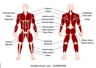
Below are two human body muscle diagrams, showing the front and the most powerful muscles in the body and those that run along the spine. Start studying muscles of the body. The eye consists of the sclera, a tough outer layer, cornea, the crystal clear curved part, the iris colored part behind the cornea, the pupil, round opening in the iris which allows light to enter. Human internal organs and human sensory system in the skin. Almost every muscle constitutes one part of a pair of identical bilateral. There are approximately 640 skeletal muscles within the typical human, and almost every muscle constitutes one part of a pair of identical bilateral muscles, found on both sides, resulting in approximately 320 pairs of muscles, as presented in this article. Now that you know how muscle tissue is made up and how muscles interact with the bones and nervous system, it helps to know where exactly these muscles reside in the body. Their main purpose is to help us to move our body parts. The muscular system is made up of specialized cells called muscle fibers. How to study muscle anatomy. Muscles allow a person to move the ear contains the smallest muscles in the body alongside the smallest bones. Cardiac muscle contracts the heart to pump blood. These muscles hold the inner ear together and are connected to the.
This is a table of muscles of the human anatomy. Small muscles arising from the back portion of hand and foot and extending to base of little finger or little toe. Below are two human body muscle diagrams, showing the front and the most powerful muscles in the body and those that run along the spine. Found only in the heart, cardiac muscle is responsible for pumping blood throughout the body. The muscles of the spine anatomy chart shows every one of the many layers of muscle in the spine and back, using.
Anatomy And Physiology Lab I On Openalg from alg.manifoldapp.org These muscles hold the inner ear together and are connected to the. Here are more details about the structure and function of each type of muscle tissue in the human muscular system. Their main purpose is to help us to move our body parts. Muscles are made up of 30% protein and 70% salt solution. They maintain posture and provide the strength for lifting and pushing. The muscular system constitutes about 45% of our total body weight. Dot pattern simple kolams collection beautiful kolams, dot sikku kolam pattern, kolam picture, kolam pattern, kolam drawing. The pectorals are located at the front of the upper body.
To get started, choose a muscle group either on the muscle chart.
Human body muscle system, the muscles of the human body that work the skeletal system, that are under voluntary control, and that are concerned with movement, posture, and balance. While cardiac muscles are exclusively found in the heart, skeletal and smooth muscle tissue. They are categorized by the muscles which they affect (primary and secondary), as well as the equipment required. Found only in the heart, cardiac muscle is responsible for pumping blood throughout the body. You are what eat infographic vector. When a muscle is activated it contracts, making itself shorter and thicker, thereby pulling its ends closer. Now that you know how muscle tissue is made up and how muscles interact with the bones and nervous system, it helps to know where exactly these muscles reside in the body. Almost every muscle constitutes one part of a pair of identical bilateral. In the diagrams below, i'll be showing muscle groups in color, with a black line to show the forms a muscle's job is to pull together the points to which its ends are attached. Small muscles arising from the back portion of hand and foot and extending to base of little finger or little toe. Muscle is a tissue in animal bodies. • small arteries and arterioles are distinguished from one another by the number of smooth muscle cell layers in the tunica media. The muscular system consists of various types of muscle that each play a crucial role in the function of the body.
You are what eat infographic vector diagram of muscles in the body. Is a tendon of the back of the leg, and the thickest in the human body.


0 Komentar